Severe flank pain can come from a passing stone, an obstructed kidney, infection, or a condition outside the urinary tract. By the end, you will know when to call a urologist the same day, when to go straight to an emergency department, what details and tests matter, and why stone size and location often matter more than pain intensity.
Key takeaways
- Fever, shaking chills, vomiting, or reduced urine with flank pain needs emergency care.
- A CT scan without contrast can show stone size, location, and urinary blockage.
- Small stones near the bladder often pass; larger or obstructing stones need urologic review.
- Severe pain without fever still needs prompt assessment when it persists or returns.
Which flank-pain symptoms make kidney stones an emergency?
A kidney stone emergency is possible when obstruction threatens kidney function or infection is trapped behind it. Flank pain with fever, shaking chills, or urinary symptoms can indicate an infected obstructed kidney; waiting for the stone to pass can allow sepsis to develop.
Go to an emergency department rather than managing this at home, especially if you have one functioning kidney or cannot pass urine.
- Call emergency services for confusion, fainting, extreme weakness, trouble breathing, clammy skin, or rapidly worsening illness. These are sepsis symptoms or signs of circulatory collapse.
- Go to the emergency department for repeated vomiting that prevents drinking, no urine or sharply reduced urine, severe pain uncontrolled by appropriate analgesia, or pain that is getting worse.
- Seek immediate assessment for pregnancy, a solitary kidney, known blockage of both ureters, or acute kidney injury, even without fever.
| Option | What it means | When it applies |
|---|---|---|
| Emergency services | Immediate transport and stabilisation | Confusion, fainting, breathing difficulty, collapse, or severe deterioration |
| Emergency department | Urgent tests, antibiotics, fluids, and drainage if needed | Fever or chills, vomiting, inability to urinate, uncontrolled pain, or suspected infected obstruction |
| Same-day urologist | Prompt specialist review without waiting for routine care | Persistent stone symptoms, ongoing obstruction, or a stone that has not passed |
| Routine appointment | Outpatient assessment | Mild, improving pain with normal urine output and no infection or kidney-risk signs |
When should you call a urologist, visit the emergency department, or book an appointment?
Severe renal colic does not automatically mean surgery, but uncontrolled stone pain needs urgent assessment. Knowing when to see a urologist for kidney stones depends on danger signs, kidney function, and whether you can drink and urinate.
| Option | What it means | When it applies |
|---|---|---|
| Same-day urologist | Urgent specialist review without waiting for a routine slot | Pain remains intolerable despite prescribed analgesia, symptoms persist, the stone has not passed, or the stone is unlikely to pass |
| Emergency department | Immediate examination, urine and blood tests, imaging, fluids, antibiotics, and urgent urologic coordination | Flank pain with fever or chills, repeated vomiting that prevents fluids, inability to urinate, pregnancy, a solitary or only functioning kidney, worsening symptoms, or suspected obstruction |
| Emergency services | Ambulance-level help when travelling is unsafe | Fainting, confusion, collapse, severe weakness, or signs of sepsis such as rapid breathing and extreme illness |
| Routine outpatient evaluation | Planned assessment and follow-up imaging | Mild or controlled pain, normal urination, no fever, and a stable condition; arrange review if the diagnosis is uncertain or the stone has not passed |
An infected obstructed kidney is an emergency, not a “wait for it to pass” problem. The hospital may start antibiotics and drain the kidney with a ureteral stent or nephrostomy; definitive stone removal usually waits until infection resolves. Kidney stone triage should favour the emergency department when you cannot reach a urologist immediately.
How does kidney-stone pain differ from other causes of flank pain?
Renal colic usually starts suddenly, builds and eases in waves, and makes you shift position because no posture feels comfortable. This kidney stone pain pattern is called colicky flank pain.
The pain often travels from the back or side toward the lower abdomen, testicle, or labia; pain radiating to groin strongly supports ureteric irritation, but it does not prove a stone.
| Pattern | Features that support it | Clues against it |
|---|---|---|
| Renal colic | Waves of severe flank pain, nausea, urinary urgency or blood in urine | Pain that stays mild and unchanged for hours |
| Muscle or spine pain | Worse with twisting, lifting, pressing the area, or a specific movement | No movement-related change |
| Appendicitis versus kidney stones | Appendicitis often settles in the right lower abdomen with appetite loss and tenderness; a stone more often moves from flank to groin | Atypical location or progressive abdominal tenderness |
Tell the clinician exactly when the pain began, whether it comes in waves, where it started, and where it moved. Report vomiting, fever or chills, urinary burning, frequency, visible blood, and your last urination.
- State whether you have had stones, urinary infections, abdominal surgery, or one functioning kidney.
- List your medicines, allergies, pregnancy status, and the painkillers and doses you have taken.
- Mention whether pain responds to medication, whether you can drink, and whether symptoms are worsening.
- If the pattern does not fit renal colic or remains unexplained, ask about alternative causes and appropriate imaging.
Which tests show whether a stone is obstructing the kidney?
A kidney stone urinalysis looks for blood, white cells, and bacteria, but a normal result does not rule out a ureteric stone. Send urine for a urine culture when white cells, fever, or infection symptoms raise concern; culture identifies bacteria and guides antibiotic choice.
| Test | What it shows | Important limitation |
|---|---|---|
| Creatinine blood test | Kidney injury or reduced filtration from obstruction | A normal value does not exclude early or one-sided obstruction |
| Non-contrast CT kidney stone scan | Stone size and position, swelling of the collecting system, obstruction, and alternative causes such as appendicitis or diverticulitis | Avoid routine radiation exposure in pregnancy; ultrasound is preferred initially |
| Ultrasound | Hydronephrosis, kidney size, and sometimes the stone | It can miss small ureteric stones or fail to show the exact blockage |
| Urinalysis and urine culture | Blood, white cells, and bacterial infection | Neither test reliably proves or excludes obstruction |
Reduced urine output, a solitary functioning kidney, bilateral obstruction, rising creatinine, or worsening kidney swelling increases urgency. An obstructed kidney with suspected infection requires urgent drainage with a ureteric stent or nephrostomy plus antibiotics; waiting for the stone to pass can lead to sepsis and kidney damage.
CT or ultrasound can also reveal non-urologic causes, including ovarian disease, aortic disease, diverticulitis, or muscle injury.
How do stone size and location determine observation or treatment?
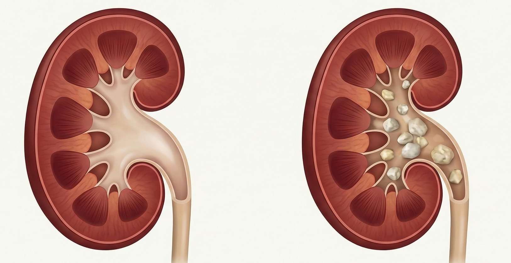
A stone 5 mm or smaller in the distal ureter has a better chance of spontaneous passage than a larger stone or one near the kidney. Stone size and passage prospects do not follow pain intensity: a tiny stone can cause severe colic, while a larger stone can cause little discomfort.
Observation is reasonable when all of these conditions apply:
- Imaging shows no infection, significant kidney impairment, or worsening obstruction.
- You can drink fluids, urinate, and control the pain with prescribed analgesia.
- The stone is small and distal rather than large or located in the proximal ureter.
- You have a follow-up plan, including imaging to confirm passage or stable position.
- An alpha-blocker is considered only for selected adults with an uncomplicated distal ureter stone, particularly when it is larger than 5 mm; it does not treat infection or dangerous obstruction.
Persistent obstruction, failure to pass on follow-up, an unlikely-to-pass stone, impaired kidney function, or pain that remains intolerable requires urologic treatment. An obstructed kidney with infection needs urgent drainage rather than further observation. Dr Zaffar Sayed Urologist can use the CT size, location, kidney function, and your medical history to select the appropriate approach.
| Option | How it works | When it fits |
|---|---|---|
| Ureteroscopy | Removes or fragments the stone through the urinary tract | Useful across many locations and sizes |
| Shock-wave lithotripsy | Breaks the stone from outside the body | Selected stones when anatomy and stone position suit targeting |
Related service
Kidney Stone Treatment Kidney stones can be as small as a grain of sand or as large–sometimes larger to an extent that it measures more than 5 cm in size. View service → |
Frequently asked questions
Which flank-pain symptoms make kidney stones an emergency?
Seek emergency care for flank pain with fever, shaking chills, confusion, repeated vomiting, very little urine, or severe weakness. An infected blocked kidney can lead to sepsis.
When should you call a urologist, visit the emergency department, or book an appointment?
Visit the emergency department for possible infection, uncontrolled pain, repeated vomiting, or reduced urine. Call a urologist promptly for persistent or recurring pain, a known stone, or a scan showing obstruction. Book an appointment for stable symptoms needing diagnosis or prevention planning.
How does kidney-stone pain differ from other causes of flank pain?
Stone pain often comes in waves, spreads toward the groin, and may cause nausea or blood in the urine. Constant pain with fever suggests infection; pain linked to movement can point to muscle or spine problems.
Which tests show whether a stone is obstructing the kidney?
A non-contrast CT scan of the urinary tract shows the stone, its size, location, and degree of obstruction. Ultrasound can show swelling of the kidney, while urine and blood tests check for infection and impaired kidney function.
How do stone size and location determine observation or treatment?
Small stones, especially those near the bladder, are more likely to pass with monitoring and pain control. Larger stones, upper-ureter stones, persistent obstruction, infection, or uncontrolled pain may require procedures such as ureteroscopy or shock-wave lithotripsy.