Blood in the urine, flank pain and urinary symptoms can come from a stone or infection, but they can also signal a tumour in the ureter or renal pelvis. You will learn how these cancers differ, which tests establish the diagnosis, how doctors classify risk and why treatment ranges from kidney-sparing procedures to removal of the kidney and ureter.
Key takeaways
- Blood in the urine needs medical assessment, even when painless or occasional.
- Smoking and previous urinary-tract cancer increase ureteric cancer risk.
- Diagnosis can include urine tests, CT urography, ureteroscopy and tissue biopsy.
- Treatment depends on tumour grade, stage, location and kidney function.
What is ureteric cancer and which symptoms need attention?
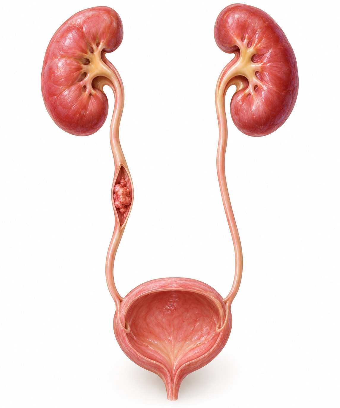
The ureters are two narrow tubes that carry urine from each kidney to the bladder. Ureteric cancer usually begins in their urothelial lining, the same type of lining found throughout the urinary tract. It is different from renal-cell cancer, which starts in kidney tissue.
Cancer can also begin in the renal pelvis, the urine-collecting area inside the kidney. These are separate starting points, but both are classified as upper-tract urothelial carcinoma.
| Site | Where it starts | Cancer category |
|---|---|---|
| Ureteric cancer | Lining of the tube between kidney and bladder | Upper-tract urothelial carcinoma |
| Renal pelvis cancer | Lining of the urine-collecting area inside the kidney | Upper-tract urothelial carcinoma |
As a tumour grows, it can narrow the ureter and slow urine drainage. This pressure can enlarge the kidney, causing hydronephrosis and, if untreated, loss of kidney function.
Common ureteric cancer symptoms include:
- Visible or microscopic blood in urine
- Flank or back pain
- Painful urination or increased frequency
- Recurrent urinary infections
- Nausea caused by obstruction
- Unexplained weight loss or fatigue in advanced disease
Painless, intermittent blood in urine still needs assessment, even when it disappears. Seek urgent care for heavy or persistent clots, inability to pass urine, fever with flank pain, severe worsening pain, vomiting, or suspected obstruction when you have one functioning kidney.
Who is at risk, and why symptoms alone cannot identify the cause
Smoking and urothelial cancer are firmly linked: tobacco chemicals reach the urine and repeatedly expose the urinary lining. Other ureteric cancer risk factors include workplace exposure to aromatic amines, previous urothelial cancer, pelvic radiation, long-term analgesic exposure and Lynch syndrome.
Lynch syndrome deserves particular attention because it raises upper-tract urothelial cancer risk. A relatively young patient, a strong family history, or several colorectal or endometrial cancers among relatives can justify genetic assessment and counselling, even without a known family mutation.
| Condition | Symptoms that can overlap | Why symptoms cannot settle the cause |
|---|---|---|
| Kidney stones | Blood in urine, flank pain, nausea | Stone-like pain can occur with obstruction from another cause |
| Urinary infection | Burning, frequency, blood, flank discomfort | Recurrent symptoms need testing rather than repeated self-treatment |
| Ureteric stricture | Slow drainage, flank pain, recurrent infection | Imaging is needed to distinguish narrowing from a tumour |
| Benign urinary problems | Frequency, urgency or microscopic blood | Benign symptoms do not exclude upper-tract disease |
A normal urine dipstick does not rule out cancer. One negative urine cytology result is not enough, because voided cytology detects high-grade disease more reliably than low-grade disease. Symptoms that disappear can return after intermittent bleeding or obstruction.
Seek evaluation when urinary symptoms recur, especially with blood in the urine, instead of repeatedly taking leftover antibiotics or painkillers. Finding the cause protects kidney function and prevents a delayed diagnosis.
How do doctors diagnose a tumour in the ureter?
Doctors usually diagnose a ureteric tumour through tests that move from urine and blood checks to imaging, bladder inspection and, when needed, tissue sampling.
1. Urine microscopy looks for red blood cells and signs of infection. A normal dipstick or a single negative result does not exclude cancer.
2. Urine culture checks whether bacteria are causing the urinary symptoms. Urine cytology looks for malignant cells and is more useful for high-grade disease than low-grade disease.
3. Blood tests measure creatinine and other kidney-function markers before treatment and before using iodinated contrast.
4. CT urography is the preferred imaging test when appropriate. It has approximately 92% sensitivity and 95% specificity, but small, flat or low-grade lesions can still be missed.
5. MRI provides an alternative when CT contrast is unsuitable. Your doctor will match the scan to your kidney function and the clinical question.
6. Cystoscopy checks the bladder because urothelial tumours can occur at more than one site, not only in the ureter.
7. Ureteroscopy directly inspects the upper urinary tract and permits targeted biopsy. A small biopsy can under-stage invasion or under-grade disease, so doctors interpret it alongside imaging, urine cytology, tumour size, focality and appearance.
Follow contrast preparation, fasting and sedation instructions exactly as provided by the treating team. Ask when you may eat, drink, take regular medicines and resume normal activity after sedation.
How is treatment chosen for ureteric cancer?
Treatment is usually chosen by classifying the cancer as low-risk or high-risk UTUC, then weighing tumour control against kidney function. Low-risk features are a single tumour under 2 cm, low-grade cytology or biopsy, no invasive appearance on imaging and no aggressive variant histology.
| Risk pattern | Usual direction |
|---|---|
| Low-risk and technically treatable | Kidney-sparing treatment |
| High-grade, invasive, multifocal, large or variant-histology disease | More extensive therapy |
| High-risk localised disease | Radical nephroureterectomy |
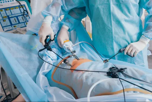
Kidney-sparing treatment uses ureteroscopy to ablate the tumour with laser or another endoscopic method. Selected distal-ureter tumours can instead undergo segmental ureterectomy, removal of that ureter segment and reimplantation into the bladder. These approaches preserve renal tissue but bring more recurrences and repeat procedures, so reliable cystoscopy, imaging and ureteroscopic surveillance is essential.
Radical nephroureterectomy removes the kidney, entire ureter and a bladder cuff. It offers the main curative operation for high-risk localised disease, but reduced renal function after surgery can prevent later cisplatin treatment.
Selected high-risk patients receive neoadjuvant cisplatin-based chemotherapy before surgery. After surgery, locally advanced or node-positive disease may receive gemcitabine plus a platinum agent; the POUT trial reported about 71% versus 46% three-year disease-free survival. A single postoperative bladder chemotherapy instillation prevents new bladder recurrences; it does not treat residual ureteric tumour.
Dr Zaffar Sayed Urologist can help patients in Mumbai discuss staging, renal function, surgical approach, chemotherapy timing and second opinions.
Related service
Uro-Oncological Management Urologic oncology is the field of medicine concerned with the research and treatment of cancers of the urinary system for both genders, as well as... View service → |
What follow-up and outlook should you expect after treatment?
Prognosis depends on stage, tumour grade, depth of invasion, lymph-node status, tumour biology, kidney function and response to treatment—not on symptoms alone. Disease-free survival describes how long cancer remains undetected after treatment; it cannot be predicted from whether blood in the urine or pain has stopped.
| Factor | Why it matters |
|---|---|
| Stage and invasion | Tumour confined to the ureter has a better outlook than cancer extending through its wall or into nearby tissue. |
| Grade and tumour biology | High-grade cells and aggressive variant histology carry greater recurrence and spread risk. |
| Lymph nodes | Clear nodes support a better outlook than cancer found in sampled nodes. |
| Kidney function | Removing a kidney can reduce treatment choices and affects long-term monitoring. |
| Treatment response | The response to chemotherapy or other treatment helps refine the outlook. |
Bladder recurrence after radical nephroureterectomy is reported in roughly 22–47% of patients. Completing ureteric cancer follow-up therefore matters even after successful surgery. Postoperative surveillance usually involves repeated cystoscopy, urine cytology and upper-tract imaging, with timing and test choice tailored to tumour risk and the treatment performed.
Take these questions to each follow-up visit:
- Was the tumour low risk or high risk?
- Were the surgical margins and lymph nodes clear?
- How has my kidney function changed?
- When is each cystoscopy or scan due?
- Which symptoms require an earlier review?
Seek urgent review for new clots, inability to urinate, fever with flank pain, vomiting or rapidly worsening pain.
Frequently asked questions
What symptoms need attention with ureteric cancer?
Blood in the urine is the main warning sign. Flank pain, painful urination, frequent urination or unexplained weight loss also need medical assessment.
Who is at risk of ureteric cancer?
Risk increases with smoking, older age, previous urothelial cancer and certain workplace chemical exposures. Symptoms alone cannot identify the cause.
How do doctors diagnose a tumour in the ureter?
Doctors may use urine tests, CT urography or other imaging, ureteroscopy and a biopsy to examine abnormal tissue and determine its grade.
How is treatment chosen for ureteric cancer?
Treatment depends on the tumour’s stage, grade, size, location, spread and your kidney function. Options can include endoscopic treatment, surgery, chemotherapy or immunotherapy.
What follow-up is needed after treatment?
Follow-up checks monitor for recurrence in the ureter, bladder or urinary tract. Your schedule depends on the cancer’s risk and the treatment received.