Bladder stones usually form when urine stays in the bladder instead of being emptied completely, although a stone can also travel down from the kidney or ureter. You will learn how to distinguish the causes and symptoms, which tests identify the problem, and when observation, endoscopic treatment or surgery is appropriate.
Key takeaways
- Incomplete bladder emptying is the main reason stones grow inside the bladder.
- Blood in urine, painful urination and interrupted flow need medical assessment.
- Tests identify the stone, its size and the cause of urine retention.
- Treat the underlying blockage to reduce the risk of recurrence.
How bladder stones differ from kidney and ureteric stones
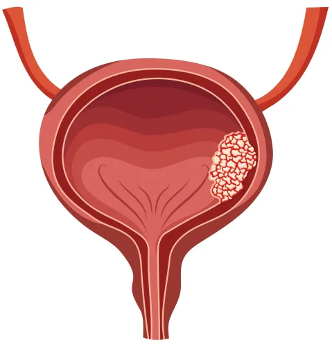
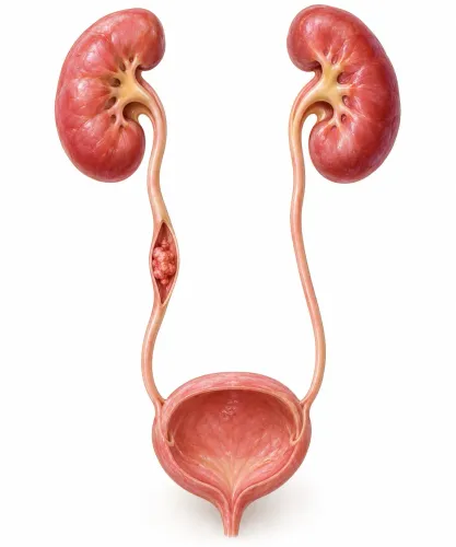
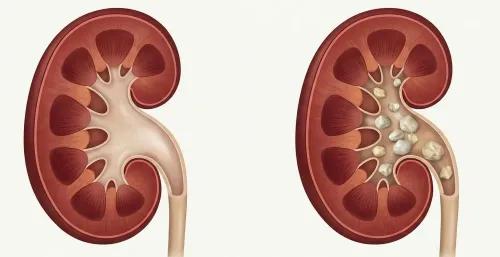
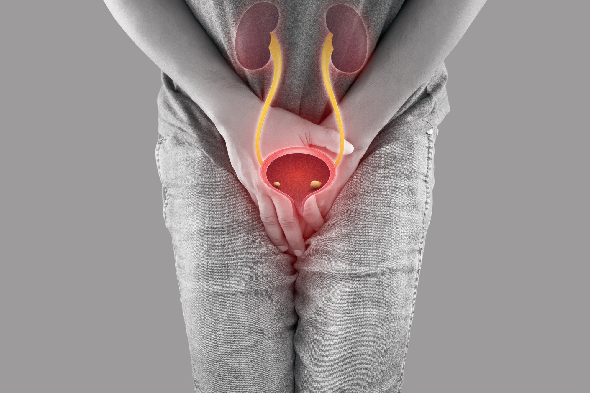
A bladder stone forms inside the bladder, usually when urine remains after urination and dissolved minerals crystallise. It may also reach the bladder when a kidney stone passes through a ureter; a stone that grows in the bladder more often signals incomplete emptying or an outlet problem.
| Stone type | Where it forms | How it travels and commonly feels |
|---|---|---|
| Bladder stones | Inside the bladder | Develop in stagnant urine or arrive from above; cause lower abdominal pain, painful urination, or interrupted flow |
| Kidney stones | Kidney collecting system | May stay in the kidney or pass downward; commonly cause flank or back pain, nausea, and blood in urine |
| Ureteric stones | Ureter, the tube from kidney to bladder | Move toward the bladder; often cause severe wave-like side or groin pain and urinary urgency |
A bladder stone is causing trouble when you notice:
- Pain or burning while urinating, especially pain that worsens at the end of the stream
- Urinary frequency, urgency, or difficulty starting
- A weak or interrupted stream that stops when the stone blocks the bladder outlet
- Lower abdominal or penile pain, sometimes triggered by movement
- Visible blood in the urine, or blood detected on testing
- Fever, chills, vomiting, inability to urinate, or rapidly worsening pain, which require urgent medical assessment
Why urine retention creates bladder stones
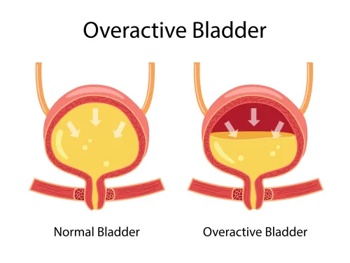
Urine does not need to stop completely for minerals to settle. Incomplete bladder emptying leaves residual urine behind; with repeated urinary retention, that stagnant pool becomes concentrated and crystals can grow around mucus, blood, bacteria or another small particle.
Common reasons include:
- BPH narrowing the bladder outlet in older men
- A urethral stricture, caused by scarring, injury or previous instrumentation
- Neurogenic bladder, when nerve disease or spinal injury weakens bladder contraction
- Pelvic organ prolapse, which can change the position of the bladder outlet
- A bladder diverticulum, where urine collects in an outpouching
- Recurrent infection, a long-term catheter or another foreign body
Age and anatomy change the likely cause. Older men face the greatest risk from prostate enlargement and outlet obstruction, while women can develop retention when pelvic organ prolapse or nerve-related bladder dysfunction prevents complete emptying.
Children need a different assessment: infection, abnormal urinary-tract anatomy, poor bladder emptying, diet and inherited or metabolic stone conditions can all contribute. Their stones are not simply an older-man obstruction problem.
A stone that migrated from the kidney or ureter follows a separate pathway. Removing that stone without correcting persistent obstruction, infection or poor bladder contraction leaves the retention problem in place, so another stone can form. Measuring post-void residual urine helps show whether the bladder is actually emptying.
Symptoms, tests and the point when treatment is needed
A clinician confirms a bladder stone with imaging, usually ultrasound or a CT scan, and may inspect the bladder with cystoscopy when the diagnosis remains uncertain. Symptoms that support the finding include painful urination, urinary frequency or urgency, an interrupted urine stream, lower-abdominal pain, or visible or microscopic blood.
- Urinalysis checks for blood, crystals and signs of infection.
- Urine culture identifies bacteria before treatment or instrumentation.
- Uroflowmetry measures how strongly urine leaves the bladder.
- A post-void residual scan measures urine left after you finish urinating.
An incidental stone does not automatically require removal. Active treatment becomes more important when it causes pain, recurrent infection, blood in the urine, urinary obstruction, retention, or progressive symptoms. A stone near the bladder outlet deserves particular attention because it can suddenly block flow.
A small, symptom-free stone may be monitored when urine drains well and there is no infection, but follow-up must address why it formed. Enlarged prostate, urethral narrowing, weak bladder contraction, a diverticulum, or a catheter can keep urine stagnant.
Removing the stone while leaving obstruction untreated can lead to recurrence, so clinicians weigh residual urine, prostate findings, stone size and location, other illnesses, and operative risk before choosing surveillance or removal.
What bladder-stone treatment involves
For most symptomatic adults, a urologist removes the stone through the urethra rather than making an abdominal incision. This endoscopic approach is called cystolithotripsy or cystolitholapaxy: the first term describes breaking the stone, while the second includes removing the resulting pieces.
| Treatment | What happens | When it applies |
|---|---|---|
| Endoscopic laser treatment | A cystoscope passes through the urethra into the bladder. Laser fragmentation breaks the stone into dust or removable pieces, which are washed out or extracted; a temporary catheter may remain afterward. | Usual option for accessible stones in adults |
| Percutaneous removal | The surgeon creates a small channel through the lower abdomen into the bladder, inserts instruments, fragments the stone and removes the pieces. | Large stones, difficult urethral access, or anatomy that makes urethral treatment unsuitable |
| Open surgery | The bladder is opened through an abdominal incision and the stone is removed directly. | Very large or numerous stones, complex anatomy, or failed or unsuitable endoscopic treatment |
Laser treatment is not complete when the stone has merely been cracked. The surgeon checks the bladder for retained fragments and addresses bleeding, infection risk and the reason urine was left behind. Catheter care means keeping the drainage bag below bladder level, avoiding kinks and reporting fever, worsening pain or blocked drainage.
If prostate enlargement or another outlet blockage caused the stone, removing it alone leaves the recurrence risk in place. Dr Zaffar Sayed Urologist can assess whether obstruction, urethral narrowing or poor bladder contraction needs treatment during the same operation or separately.
Preventing recurrence and preparing for a urology consultation
Reduce recurrence by treating the reason urine remained in the bladder, not by relying on extra water alone. Drink enough to produce pale urine unless a clinician has restricted your fluids, avoid routinely delaying urination, and follow the prescribed plan for constipation, infection, catheter care, or prostate and urethral obstruction.
Ask for stone analysis; its composition can distinguish infection-related, metabolic, foreign-body, and migrated stones.
At the consultation, take your scan reports, medication list, culture results, catheter history, and a record of urinary symptoms. Children need assessment of urinary-tract anatomy, bladder emptying, diet, infection, and inherited or metabolic stone risk rather than an adult-style prostate evaluation.
- What was my post-void residual urine, and has it fallen after treatment?
- What measured my urine flow, and does the result show continuing obstruction?
- If I have prostate enlargement, urethral narrowing, prolapse, or a diverticulum, has that cause been treated or only the stone removed?
- Could neurogenic bladder, a catheter, or another foreign body be keeping urine in place?
- If I have recurrent urinary infection, was a urine culture obtained, and what changed to prevent another infection?
- What follow-up test will confirm that emptying remains adequate?
| Finding or cause | Question to ask | Evidence of control |
|---|---|---|
| Prostate or urethral obstruction | Is the outlet now open? | Better flow and lower residual |
| Neurogenic bladder | Is a drainage plan needed? | Safe residual and scheduled review |
| Infection-related stone | What did stone analysis and culture show? | Cause-specific treatment and no repeat infection |
Frequently asked questions
How do bladder stones differ from kidney and ureteric stones?
Bladder stones form in the bladder, while kidney stones form in the kidneys and ureteric stones travel through the tubes from the kidneys to the bladder.
Why does urine retention create bladder stones?
Urine left in the bladder becomes concentrated, allowing dissolved minerals to crystallise and grow into stones. Retention often results from an enlarged prostate, urethral narrowing, nerve-related bladder dysfunction or another outlet problem.
What symptoms and tests indicate a bladder stone?
Symptoms include painful urination, blood in the urine, frequent urination, lower abdominal pain and an interrupted urine stream. Evaluation can include urine testing, ultrasound, imaging and cystoscopy.
When is bladder-stone treatment needed?
Treatment is needed when the stone causes symptoms, infection, bleeding, obstruction or fails to pass. The underlying cause of incomplete emptying also needs attention.
What does bladder-stone treatment involve?
A urologist may remove a small stone through the urinary passage or break it into smaller pieces with an endoscopic procedure. Larger stones require a different removal approach, followed by treatment of the cause.